Friday, 14 October 2016

CIE XYZ:


The basic CIE colour space, or colour model, is based on a 'Standard Observer and 'Standard Illuminants' (D50, D65, etc.). This is a numerical model of colour sensitivity based on research commenced in the 1920s on a sample of people with normal colour vision. It is a 'universal colour space' representing the colour spectrum visible to the 'average human'. The light-sensitive retina at the back of the eye has three types of receptors near the centre, known as cones. They are sensitive to the three primaries, 'red, green and blue'. The CIE XYZ tristimulus values are assigned to the red, green and blue curves respectively. These approximate to the cones in the eye. The relative response of each is plotted on a diagram against the wavelength in nanometers. The eye also has rods, outside of the retina's centre, which are sensitive to low-wavelength light and which only operate at low levels of illumination. There are two axes: vertical and horizontal.

The vertical axis represents Relative Response 0 - 2.0 (shown here) or Reflective Intensity 0 - 120% (not shown).
The horizontal axis represents Wavelength in nanometers, usually from about 380 to about 720nm.
It should be emphasized that this is a 'device-independent' colour space in which each primary colour (X,Y,Z) is always constant, unlike  RGB which varies with every individual device (monitor, scanner, camera, etc.). XYZ is typically used to report the spectral response of a sample measured by a colorimeter or a spectrophotometer. A colorimeter may contain as few as three sensors, one each for red, green and blue, (or X, Y and Z), and will typically be used for display calibration and profiling. A spectrophotometer will report the entire spectral response at frequent intervals along the spectrum, say every 10 nanometres, and will typically be used to measure printed sheets to control a press or create an ICC profile.
While CIE XYZ is used to report colour from measuring instruments, it is not so useful for humans to describe colour. Another use is as the Profile Connection Space (PCS) within an ICC profile, where it may be used instead of CIE Lab.
You may notice that the Y ('green curve') covers the widest wavelength. This corresponds to the overall human visual response to all colours, or lightness. It is therefore also used to indicate luminance ('lightness').

CIE L*a*b*: In such case the vertical L* axis represents Lightness, ranging from 0-100.  The other (horizontal) axes are now represented by a* and b*. These are at right angles to each other and cross each other in the centre, which is neutral (grey, black or white). They are based on the principal that a colour cannot be both red and green, or blue and yellow. 
      The a* axis is green at one extremity (represented by -a), and red at the other (+a). The b* axis has blue at one end (-b), and yellow (+b) at the other. 
The centre of each axis is 0. A value of 0, or very low numbers of both a* and b* will describe a neutral or near neutral. In the case of paper, the white point in terms of a* and b* is usually carried through to the black, being gradually reduced towards '0'.
In theory there are no maximum values of a* and b*, but in practice they are usually numbered from -128 to +127 (256 levels).
The CIE Lab colour model encompasses the entire spectrum, including colours outside of human vision. CIE Lab is extensively used in many industries apart from printing and photography. Its uses include providing exact colour specifications for paint (including automotive, household, etc.), dyes (including textiles, plastics, etc.), printing ink and paper. 


CIE Color Systems

CIE Color Systems The CIE, or Commission Internationale de l’Eclairage (translated as the International Commission on Illumination), is the body responsible for international recommendations for photometry and colorimetry.

In 1931 the CIE standardized color order systems by specifying the light source (or illuminants), the observer and the methodology used to derive values for describing color.

The CIE system characterizes colour by a luminance Y and two colour coordinates x and y which specify the point on the chromatic diagram. This system offers more precision in colour measurement than do the Munsell system because the parameters are based on the spectral power distribution of the light emitted from a coloured object and is factored by sensitivity curves which have been measured for the human eye.

Based on the fact that the human eye has three different types of color sensitive cones, the response of the eye is best described in terms of three "tristimulus values". However, once this is accomplished, it is found that any color can be expressed in terms of the two color coordinates x and y.
The colors which can be matched by combining a given set of three primary colors (such as the blue, green, and red) are represented on the chromaticity diagram by a triangle joining the coordinates for the three colors.

The diagram given below represents the  mapping of human color perception in terms of two CIE parameters x and y. The spectral colors are distributed around the edge of the "color space" as shown, and that outline includes all of the perceived hues and provides a framework for investigating color.
The CIE Color Systems utilize three coordinates to locate a color in a color space. These color spaces include:

• CIE XYZ
• CIE L*a*b*
• CIE L*C*h°
Scales for Measuring Colour:
There are two important scales for measuring colour. These are:
      Munsell Scale
      CIE colour system

Munsell Scale:

In 1905, artist Albert H. Munsell originated a color ordering system — or color scale — which is still used today. The Munsell System of Color Notation is significant from a historical perspective because it’s based on human perception. Moreover, it was devised before instrumentation was available for measuring and specifying color. This system assigned numerical value to the three properties of the colour-Hue, Chroma and value. The Munsell color system match colors to a set of standard samples. The Munsell system divides hue into 100 equal divisions around a color circle and circle is distorted by assigning a unit of radial distance to each perceptable difference in saturation (called units of chroma). Since there are more perceptable differences for some hues, the figure will bulge outward to 18 values for some hues compared to only 10 for another. Perpendicular to the plane formed by hue and saturation is the brightness scale divided into a scale of "value" from 0 (black) to 10 (white). A point in the color space so defined is specified by hue, value, and chroma in the form H V/C.

The MUNSELL system is a collection of color samples for comparison, with adjacent samples based upon equal perceived differences in color.

Munsell saw that full chroma for individual hues might be achieved at very different places in the color sphere. For example, the fullest chroma for hue 5RP (red-purple) is achieved at 5/26.

Another color such as 10YR (yellowish yellow-red) has a much shorter chroma axis and reaches fullest chroma at 7/10 and 6/10:

Instrumentation for colour measurement:
As colour is perception, it cannot be directly measured; however we can measure and subsequently calculate certain factors which are responsible for producing this sensation of color. The quantification of the color properties of textile materials is of great economic value in industry and instruments are employed to some degree in almost every textile operation involved in textile coloration.

Color instrumentation has experienced a tremendous advancement in technology during the past few decades. The first devices for measuring color were absorptiometers which were used to determine by visual inspection whether two solutions were of equal color. This is very similar to holding two glass cylinders of dye solution up to a light and judging whether they are of equal strength and shade, except that the absorptiometer provided a method of adjusting the thickness or path width so that this change in width could be read from a scale. In measuring reflected light from opaque materials such as textiles, the first instruments were reflectometers developed around 1915-1920. 
Perception of colour: It involves a series of events which are interdisciplinary in nature. Perception of colours includes source of light, object that is illuminated and eye and brain that perceive the colour.
                                                   Object
Metamerism: It is a phenomenon observed when two specimens appears to have the similar colour under one set of viewing conditions, but different under another. The change in viewing conditions refers to change in source, observer or geometry of the observation.
Types of metamerism
         illuminant metamerism
         observer metamerism
         geometry metamerism
Illuminant metamerism
When the colour of two specimens matches under one illuminant but not with another illuminant, it is said to be illuminant metamerism.


Observer metamerism
When colour of two objects appears to match to one observer but not to the other, it is said to be observer metamerism. In this case observer may not be colour blind but the spectral sensitivity of colour receptors of one observer may be slightly different than the other.
Geometry metamerism
Two objects which match in one arrangement of illumination, sample and observer may mismatch by altering the positions. 

MEASUREMENT OF COLOUR
Colour is perception and sensation experienced caused by light reflected from or transmitted through objects. If you have some coloured object, it means it has certain reflectance characteristics—the patterns of light wavelengths that are reflected and absorbed—that are physical properties of object. However, colour is our subjective perception of the wavelengths of light that end up bouncing off the object and onto our retina. Photoreceptors in the retina begin the process by selectively responding to different wavelengths. A single type of photoreceptor alone cannot accomplish color vision since it cannot distinguish between enough wavelengths. We overcome this difficulty by using three different types of cone photoreceptors to code for color, a concept known as trichromacy. One of the key observations in developing the trichromatic theory of color vision was that lights can be added together to form mixtures that look identical to other, single light wavelengths. Another activity illustrates additive and other forms of color mixing.
Human Visual System

The human visual system consists of two functional parts, the eye and (part of the) brain. The brain does all of the complex image processing, while the eye functions as the biological equivalent of a camera.


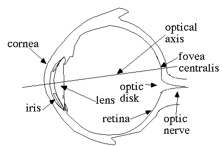
What our eyes perceive of a scene is determined by the light rays emitted or reflected from that scene. When these light rays are strong enough (have enough energy), and are within the right range of the electromagnetic spectrum (about 300 to 700 nm), the healthy eye will react to such a ray by sending an electric signal to the brain through the optic nerve. When a light ray hits the eye, it will first pass through the cornea, then subsequently through the aqueous humor, the iris, the lens, and the vitreous humor before finally reaching the retina. The cornea is a transparent protective layer, which acts as a lens and refracts the light. The iris forms a round aperture that can vary in size and so determines the amount of light that can pass through. Under dark circumstances the iris is wide open, letting through as much light as possible. In normal daylight, the iris constricts to a small hole. The lens can vary its shape to focus the perceived image onto the retina. In the retina, the light rays are detected and converted to electrical signals by photoreceptors. The eye has two types of photoreceptors: rods and cones, named after their approximate shape. The rods are abundant, about 100 million in a human eye, and spread evenly about the retina, except at the fovea, where there are almost none. The fovea is the area of the retina where our vision is sharpest. There are much fewer cones, about 6 to 7 million, which are mainly located around the fovea, but can be found in a low density in the entire retina. No photoreceptors are found at the point where the optic nerve attaches to the eye (the so-called blind spot), so we cannot perceive anything there. Since rods are more responsive to light than cones we can identify three types of vision, depending on the amount of light that reaches the eye. Under dark circumstances, practically only the rods are active. Since rods cannot discriminate colors, we perceive only shades of grey. We call this scotopic or night vision. Under daylight circumstances, the cones are most active, and we experience photopic or day vision. In dimly lighted circumstances there is an intermediate stage where both rods and cones are active called mesopic vision. We are able to distinguish colors because there are three distinct types of cones, each sensitive to a different band of the electromagnetic spectrum.

Wednesday, 30 December 2015

My Book on "Pretreatment of Textile material for dyeing and Printing" released today by Textile Commissioner of India during 56th Joint Technological Conference, Coimbatore.